MIT Research Doubles the Depth Limit of Metabolic Imaging
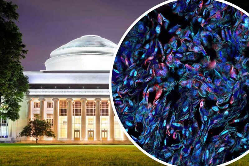
Researchers at MIT have developed an innovative method to improve the study of living cells by illuminating deeper into the analyzed tissue. This metabolic imaging technique refines disease progression assessment and treatment responses. The technique uses laser light to study living cells. Classified as “label-free” imaging, it does not require cells to be stained prior to imaging. This means images of living tissue can be better captured and monitored.
While metabolic imaging is an established non-invasive medical imaging, traditional methods have not been able to penetrate deeply enough into tissue because light scatters when shining onto tissue, reducing resolution.
What makes this new process unique is the way engineers have been able to customize the laser light using a fiber bender device. This fiber bender is paired with a multimode fiber and precisely modulates the light propagation by adaptively changing the shape of the fiber. This manipulates the intensity of the laser for enhanced deep-tissue imaging.
Tests proved that researchers could penetrate beyond 700 micrometers into biological tissue compared to previous methods which enabled only around 200 micrometers. Cells can now be imaged at multiple levels, delivering more detailed information on the metabolic state inside a living organoid while it continues to grow.
Scientists at MIT state that the new multimodal metabolic imaging method is well-suited for demanding imaging applications like cancer research, tissue engineering, drug discovery, and the study of immune responses.
Full details of the project are published on MIT’s website.